
Fig. 3.
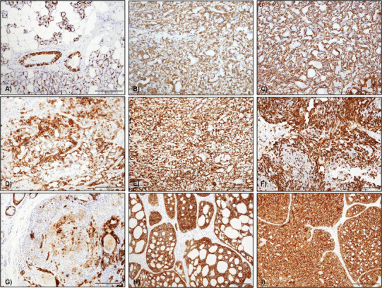
CK14 staining in salivary gland tumours a in normal tissue CK14 staining was mainly seen as a cytoplasmic staining of myoepithelial cells surrounding acini and in some basal cell in ducts, b CK14 in ACC was variable with one case showing diffuse abluminal staining, c CK14 in one SC with diffuse cytoplasmic staining of abluminal cells, d CK14 staining in PA was predominantly seen in the cytoplasm of most tumour cells, but plasmacytoid cells were negative, e CK14 expression in CA ex-PA was mainly seen in the cytoplasm of the abluminal type cells, f CK14 staining in myoepithelioma was variably positive in the cytoplasm of the neoplastic myoepithelial cells, mainly spindle cells, g CK14 staining in MC was focal with cytoplasmic staining of scattered tumour cells, i CK14 staining in AdCC (cribriform variant) and j PAC—diffuse staining was seen throughout the tumour (original magnification ×20)