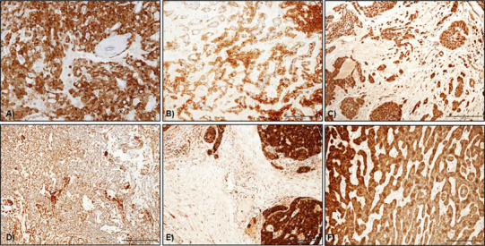
Fig. 4.

S100 staining in salivary gland tumours. a Nuclear and cytoplasmic staining in myoepithelial cells in PA whereas luminal cells were largely negative, b S100 expression in Ca ex-PA with diffuse staining in the cytoplasm of luminal, abluminal and scattered stromal cells, c myoepithelioma showing diffuse cytoplasmic S100 staining in the neoplastic spindle cells, d S100 staining in MC with strong cytoplasmic staining in the plasmacytoid cells, e PAC showed diffuse staining in almost all the tumour cells (original magnification ×20)
