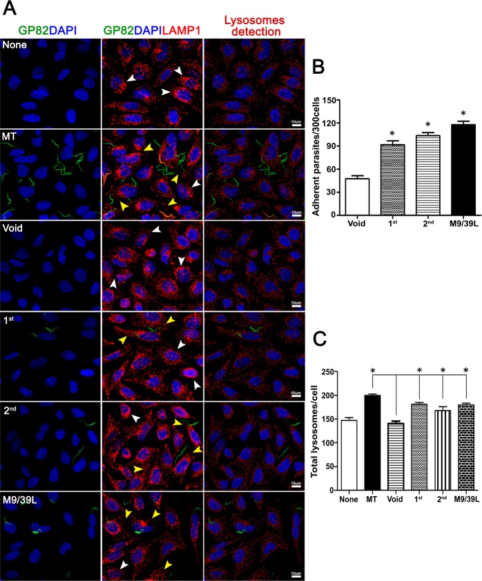
Figure 5.
T. cruzi epimastigotes expressing GP82 bind to HeLa cells and induce lysosome mobilization. (A) HeLa cells seeded on glass coverslips were incubated with transfected parasites at MOI 20:1 for 1 h at 37 °C in 24-wells plates. Wells were washed with PBS to remove unbound parasites and were fixed with 4% PFA, permeabilized with 0.1% saponin (PGN-saponin) and incubated for 1 h with mouse mAb 3F6 and rabbit mAb anti-human Lamp1 diluted in PGN-saponin. Coverslips were washed and incubated for 1 h with 2 µg/mL of anti-mouse IgG conjugated to Alexa Fluor 488 and anti-rabbit IgG conjugated to Alexa Fluor 568 containing 1 µg/mL DAPI. Samples, mounted on microscopic slides using Prolong Gold, were analysed on Leica TCS SP8 Confocal Laser Scanning Platform using Leica Application suite (LAS) and Imaris (Bitplane) software packages. Lysosome detection was performed as described elsewhere47 None: cells incubated in culture media devoid of parasites; MT: wild-type metacyclic trypomastigotes; Void: epimastigotes transfected with empty pTEX plasmid; 1st: transfected parasites carrying the pTEX-1st construct; 2nd: epimastigotes transfected pTEX-2nd construct; M9/39L: transfected parasites carrying the pTEX-M9/39L construct. White arrowheads: perinuclear lysosomes. Yellow arrowheads: lysosome scattering induced by transfected parasites. Bar: 10 μm. (B) Epimastigotes expressing GP82 were incubated with HeLa cells at MOI 20:1 for 1 h at 37 °C in 24-wells plates containing DMEM medium. After washings with PBS to remove unbound parasites and fixation in Bouin solution followed by Giemsa staining, the coverslips were mounted onto microscopic slides and the number of cell-adherent parasites recorded by microscopy. The results correspond to the mean ± SD of parasites in 300 cells counted in triplicate (* p < 0.05). (C) Quantification of lysosome mobilization/scattering from panel A analysed by lysosome counting algorithm. Bars correspond to triplicates indicating mean ± SD of 10 different microscopic fields (≥300 cells) observed with a 63× objective (* p < 0.05).

