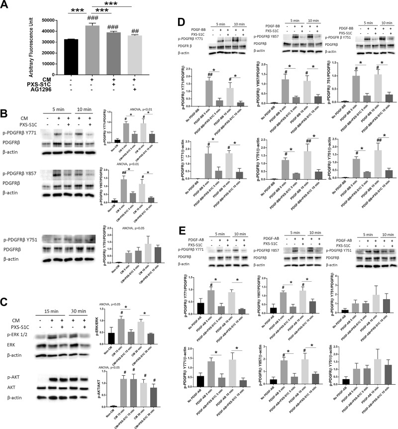
Fig. 6. HSC3 oral tumor cell-derived LOXL2 stimulates oral fibroblast proliferation and cell signaling in collaboration with PDGF-AB.
LOXL2 inhibitor PXS-S1C attenuates HSC3 CM-stimulated human oral fibroblast (a) proliferation, (b) phosphorylation of PDGFRβ at the Y771 and Y857 but not Y751 residues, and (c) ERK activation, but not AKT. a Human gingival fibroblast proliferation was reduced after 24-h treatment with PXS-S1C (1 µM) or AG 1296 (5 µM) in the HSC3 CM as determined by the CyQUANT assay. Data are means SEM. This experiment was done three times independently with six replicate samples. ANOVA, p < 0.0001, Dunnett’s multiple comparisons test, ***p < 0.0001 indicates significant difference between treated groups; while ##p < 0.001, ###p < 0.0001 indicate significant differences from non-CM group. b and c Gingival fibroblasts were serum depleted and then treated with non-CM, and CM with and without PXS- S1C (1 µM) and cell layer protein samples were subjected to western blot. Data are means ± SEM. The experiment was performed with three times independently with primary human gingival fibroblasts isolated from three different donors. Representative blots are shown. Data from all three experiments were subjected to quantitative analyses. Sidak’s multiple comparison test, *p < 0.05 indicates a significant difference from PXS-S1C treated group. Dunnett’s multiple comparison test, #p < 0.05, ##p < 0.001 indicate significant differences from Non-CM group. d PXS-S1C attenuates PDGF-BB stimulated phosphorylation of all three PDGFRβ phosphorylation sites Y771, Y857 and Y751, and AKT activation in oral fibroblasts. Gingival fibroblasts were serum depleted and then treated with no PDGF-BB, and PDGF-BB (10 ng/ml) with and without PXS-S1C (1 µM). The protein samples were subjected to western blot. Data are means ± SEM. The experiment was done with three times independently with primary human gingival fibroblasts isolated from three different donors. Representative blots are shown. Data from all three experiments were subjected to quantitative analyses. Sidak’s multiple comparison test, *p < 0.05, **p < 0.001, and ***p < 0.0001 indicate difference from PXS-S1C-treated group. Dunnett’s multiple comparison test, #p < 0.05, ##p < 0.001, ###p < 0.0001 indicate difference from No PDGF group. e PDGF-AB mimics the effects of HSC3 cell CM on oral fibroblasts in phosphorylation of PDGFRβ. Gingival fibroblasts were serum starved and then treated with no PDGF-AB, and PDGF-AB (10 ng/ml) with and without PXS-S1C (1 µM). The protein samples were subjected to western blot. Data are means ± SEM. The experiment was done with three times independently with primary human gingival fibroblasts isolated from three different donors. Representative blots are shown. Data from all three experiments were subjected to quantitative analyses. Sidak’s multiple comparison test, *p < 0.05 indicates difference from PXS-S1C-treated group. Dunnett’s multiple comparison test, #p < 0.05 indicates difference from No PDGF group

