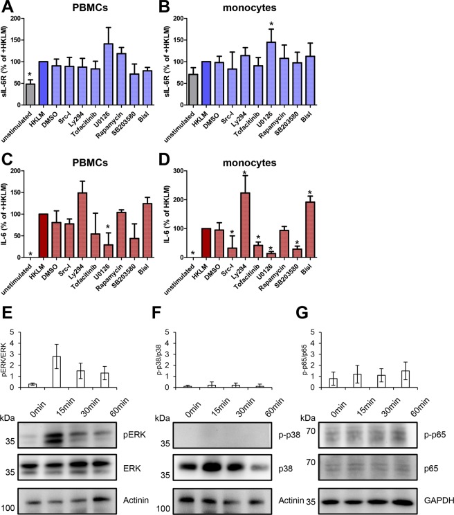
Figure 5.
Stimulation of TLR2 results in the activation of Extracellular-signal Regulated Kinase (ERK) cascade which regulates IL-6 and sIL-6R levels differentially. (A) PBMCs were isolated from plasma-free blood and were pre-incubated with the signaling pathway inhibitors Src-I (targeting the kinase Src), Ly294 (PI3K), tofacitinib (Jak1/2), U0126 (ERK), rapamycin (mTOR), SB203580 (p38/MAPK) and BisI (targeting PKC) for 90 min. Afterwards, TLR2 agonist HKLM (108 cells/ml) was added and the cells were incubated for further 24 h. The supernatants were collected and the amounts of sIL-6R were determined via ELISA. (B) Monocytes were isolated from PBMCs using the antibody-based magnetic cell separation. Stimulation of monocytes and analysis was performed as described in panel A. (C,D) The experiments were performed as described in panels A and B, but the amounts of IL-6 were determined via ELISA. Data shown are the mean ± SEM from four independent experiments (n = 8). Data were analyzed by one-way ANOVA followed by Dunnett’s Multiple Comparison test. Statistical significance compared to the HKLM-treated cells is indicated. (E–G) PBMCs were isolated from plasma-free blood, resuspended in serum-free RPMI and were starved for 2 h. The cells were stimulated with the TLR2 agonist HKLM (108 cells/ml) for 0, 15, 30 and 60 min and lysed afterwards. The indicated proteins were analyzed by Western blotting. Quantification is shown in the histograms above the Western blots. Data are representative of three independent experiments.

