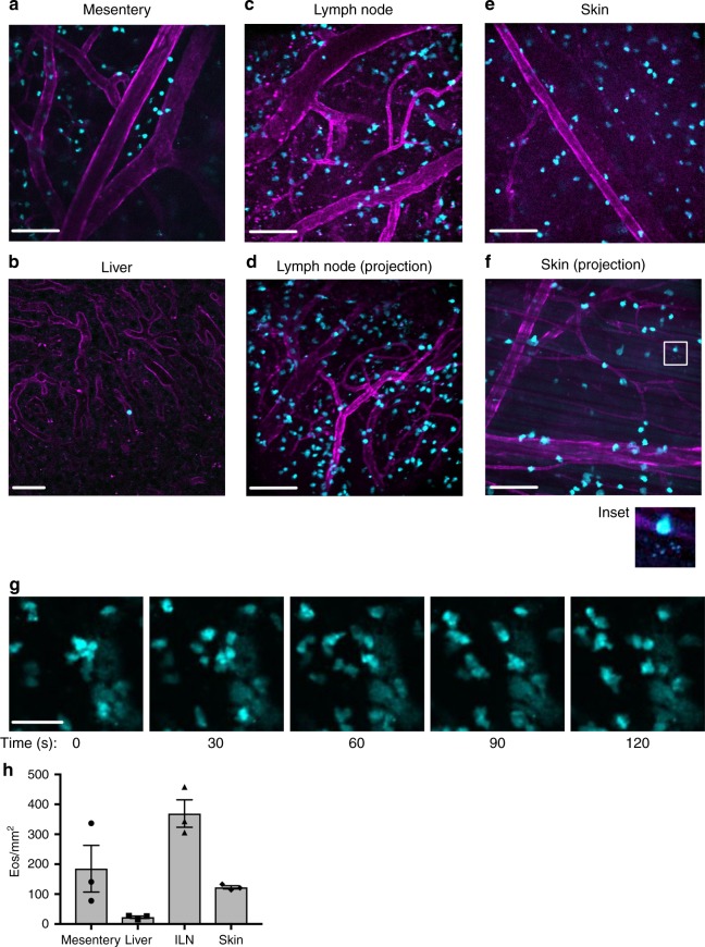
Fig. 2.
Tissue-resident eosinophils can be visualized in real time under baseline conditions in multiple organ systems using intravital microscopy. EoCRE+/−/GFP+/− mice were anesthetized and the specified organs were prepared for intravital microscopy as described in the “Methods.” Eosinophils express green fluorescent protein (cyan) and the vasculature is labeled with an anti-CD31 antibody conjugated to Alexa-594 (magenta). Representative images from the mesentery (a), liver (b), inguinal lymph node (c, d), and skin (e, f) were captured from video sequences. d and f are maximum intensity projection images generated using the optical sections from the z-stack of the tissue. Inset in f shows small punctate structures consistent with free granules. g Eosinophils from the lymph node are shown over a 90-s time frame. The full 6-min video is shown as supplementary video 1. Images are representative of between three and five experiments. Data in h are mean ± SEM; n = 3 independent experiments. Scale bars in a and c–f are 100 μm, scale bar in b is 50 μm and scale bar in g is 40 μm

