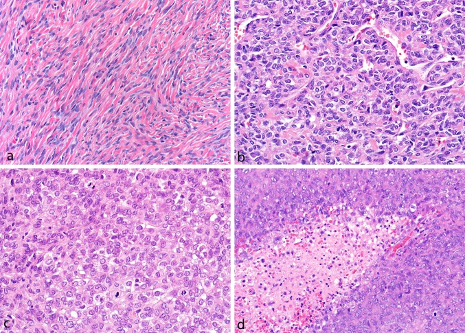
Fig. 1.
Tumors classified as solitary fibrous tumor contained uniform ovoid-to-slightly spindled-shaped cell deposited in a collagenized background and arranged around branching and hyalinized blood vessels (a), while hemangiopericytomas were highly cellular tumors composed of predominantly round cells in a sheet-like pattern (b) with less prominent vasculature often showing high mitotic rates (c) and necrosis (d)

