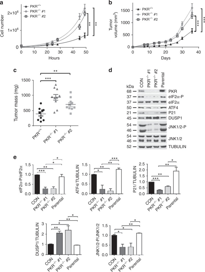
Fig. 2.
Cell-autonomous anti-tumor effects of PKR in NEU breast tumor cells. a Proliferation rates of 2 independent clones deleted for PKR by CRISPR (PKR−/−) compared with the proliferation of proficient NEU tumor cells (PKR+/+). Data is the average of 3 biological replicates. **p < 0.01. b, c NEU PKR+/+ and PKR−/− tumor cells (5 × 10⁵) were implanted in 5 female SCID mice, two injections per mouse (n = 2 × 5 = 10). Tumor volume (mm3) was monitored for the indicated days (b) and tumor mass (mg) was assessed at the experimental endpoint (c). Data represent mean ± SEM. **p < 0.01; ***p < 0.001. d Immunoblotting of NEU PKR+/+ and PKR−/− tumor cells for the indicated proteins. Protein extracts from the parental mouse NEU tumor cells, which were used for the generation of PKR−/− tumor cells by CRISPR, were included as an additional control. e Quantification of blots in d from 3 biological replicates. Data represent mean ± SEM *p < 0.05, **p < 0.01, ***p < 0.001

