Figure 4.
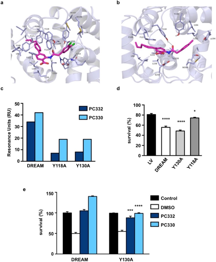
Identification of the binding site. Molecular docking model of IQM-PC332 (a) and IQM-PC330 (b) in complex with DREAM. Amino acids within 4 Å of the ligand are shown; yellow dashed lines indicated hydrogen bonds. For clarity non-polar hydrogens are not shown. (c) Direct binding of IQM-PC332 and IQM-PC330 in SPR assays to immobilized wtDREAM compared to Tyr118Ala and Tyr130Ala DREAM mutants. (d) Comparative response of sensitized STHdhQ111/111 cells to H2O2-induced oxidative stress after overexpression of wild type, Tyr118Ala or Tyr130Ala DREAM. One-way ANOVA with Holm-Sidak’s multiple comparison test (n = 6). ****p < 0.0001, *p < 0.05 vs empty vector (LV). (e) Effect of vehicle (DMSO), 130 nM IQM-PC332 or 40 nM IQM-PC330 in wild type DREAM- (DREAM) or Tyr130Ala DREAM-sensitized STHdhQ111/111 cells (Y130A). Control bar represents DREAM- or Tyr130Ala DREAM sensitized STHdhQ111/111 cells non-exposed to H2O2, respectively. The concentrations of IQM-PC compounds used in these experiments correspond to the minimum concentration able to fully reverse the effect of H2O2 exposure. Non-parametric Mann-Whitney t-test *p < 0.05 vs corresponding compound in wild type DREAM sensitized STHdhQ111/111 cells.

