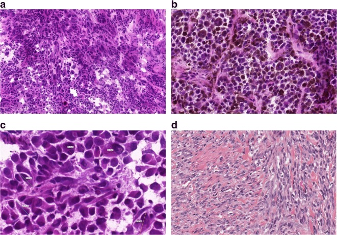
Fig. 1.
Histological appearance of melanomas. a A non-pigmented tumor composed of malignant melanocytes infiltrated by lymphocytes b A pigmented tumor producing melanin mostly composed of epithelioid shaped cells (10 ×). c Variable sizes of tumor cells are noted in a tumor; multinucleated cells can also be identified. d Spindle cell melanoma displaying fascicules of elongated melanocytes

