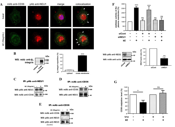
Fig. 4.
Validation of the interaction between NEU1 and CD36 in human macrophages and functional consequences on the sialylation level of CD36. a Colocalization of NEU1 and CD36 at the cell surface of macrophages differentiated from THP-1 cells, and stimulated, or not, with kE (50 µg/mL, 1 h), by confocal microscopy acquisitions. Areas of colocalization at the plasma membrane are indicated by white arrows. Images are representative of two independent experiments. b Left panel: distribution of the β2-integrin between the cytosol and crude membrane fractions (30 µg each) of macrophages by Western blot using a mouse monoclonal anti β2-integrin (1/500). The figure is representative of three independent experiments. Right panel: blots quantification by densitometry analysis. Results are expressed as mean ± SEM of three independent experiments and statistical analysis was performed by Student’s t test (***p < 0.001). c NEU1 was immunoprecipitated with a rabbit polyclonal anti-NEU1 antibody from crude membrane preparations of macrophages and co-immunoprecipitation of CD36 was monitored by Western blot using a mouse monoclonal anti-CD36 antibody. The figure is representative of three independent experiments. d CD36 was immunoprecipitated with a mouse monoclonal anti-CD36 antibody from crude membrane preparations of macrophages and co-immunoprecipitation of NEU1 was monitored by Western blot using a rabbit polyclonal anti-NEU1 antibody (1/500). The figure is representative of two independent experiments. e CD36 was immunoprecipitated with a mouse monoclonal anti-CD36 antibody from whole lysate of macrophages and co-immunoprecipitation of NEU3 was monitored by Western blot using a rabbit polyclonal anti-NEU3 antibody (1/500). The figure is representative of three independent experiments. f Up panel: sialidase activity at the plasma membrane of adherent macrophages was measured using 400 µM of 2′-(4-methylumbelliferyl)-α-d-N-acetylneuraminic acid substrate in 20 mM CH3COONa (pH 6.5) before and after incubation with kE (50 µg/mL, 2 h). Macrophages were either non-transfected or transfected with 50 nM negative control siRNA (siCont) or NEU1 siRNA (siNEU1). Results are expressed as mean ± SEM of three to nine independent experiments and normalized to the control (non-transfected, without kE). Statistical analysis was performed by one-way ANOVA followed by a Dunnett’s multiple comparisons test (***p < 0.001; ns non-significant). Down panel: expression level of NEU1 in macrophages transfected with 50 nM negative control siRNA (siCont) or NEU1 siRNA (siNEU1) and monitored by Western blot using a rabbit polyclonal anti-NEU1 antibody. The blot is representative of three independent experiments (left). Blots were quantified by densitometry analysis, and results expressed as mean ± SEM of three independent experiments and normalized to negative control siRNA (siCont). Statistical analysis was performed by Student’s t test (**p < 0.01. g SNA pulldown of crude membrane preparations of macrophages incubated, or not, with kE (50 µg/mL), V14 + kE (molar ratio 2:1) or V14 peptide alone for 1 h at 37 °C. For each condition, equal amount of proteins was used. The amount of sialylated CD36 recovered after SNA pulldown was evaluated and quantified as depicted in Fig. 3F. The sialylation level of CD36 was normalized to the respective control (w/o or V14) and results expressed as mean ± SEM of three to five independent experiments. Statistical analysis was performed by Student’s t test (*p < 0.05; ns non-significant)

