Fig. 2.
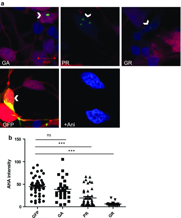
Expression of arginine-rich DPRs suppresses translation in human iPSC-derived MNs. a iPSC-MNs transfected to express DPRs (GFP-GA100, GFP-PR100, GFP-GR100) or GFP alone (arrow heads, green) were treated with AHA and newly synthesised proteins labelled with alkyne-tagged Alexa 555 (red), cells were counterstained with DAPI (blue). iPSC-MNs treated with 100 µM of anisomycin and AHA for 2 h were used as a negative control for AHA imaging (+Ani) and did not show incorporation of AHA (red). b AHA intensity was measured in DPR- or GFP-positive cells. Two inductions of control iPSC-MNs were nucleofected, from which n = 44, 28, 43, 15 for GFP-, GA-, PR- or GR-positive iPSC-MNs, respectively, were analysed. Error bars are mean ± SEM. Kruskal–Wallis test and Dunn’s Multiple comparison test, ***P < 0.001, ns indicates not significant

