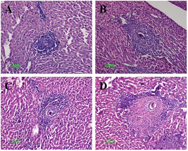
Figure 3.

Hematoxylin and eosin staining of Schistosoma egg granulomas in the livers of mice in the four infected groups. Representative photos are shown for mice in the infected-control (A), anti-cytotoxic T-lymphocyte antigen-4 monoclonal antibody (anti-CTLA-4 mAb) (B), fatty acid-binding protein (FABP) (C), and combination group (D). Scale bar, 100 μm.
