
Fig. 1.
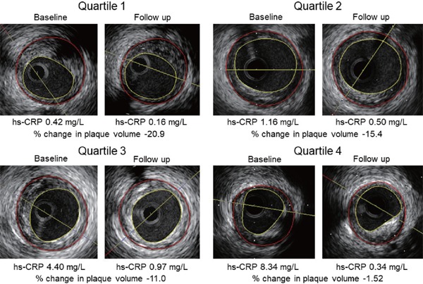
Representative intravascular ultrasound images of coronary plaque regression after lipid- and blood pressure-lowering therapy across the baseline high-sensitivity C-reactive protein (hs-CRP) level quartiles. The percentage change in the plaque volume rose progressively with increasing the baseline hs-CRP.