FIGURE 2.

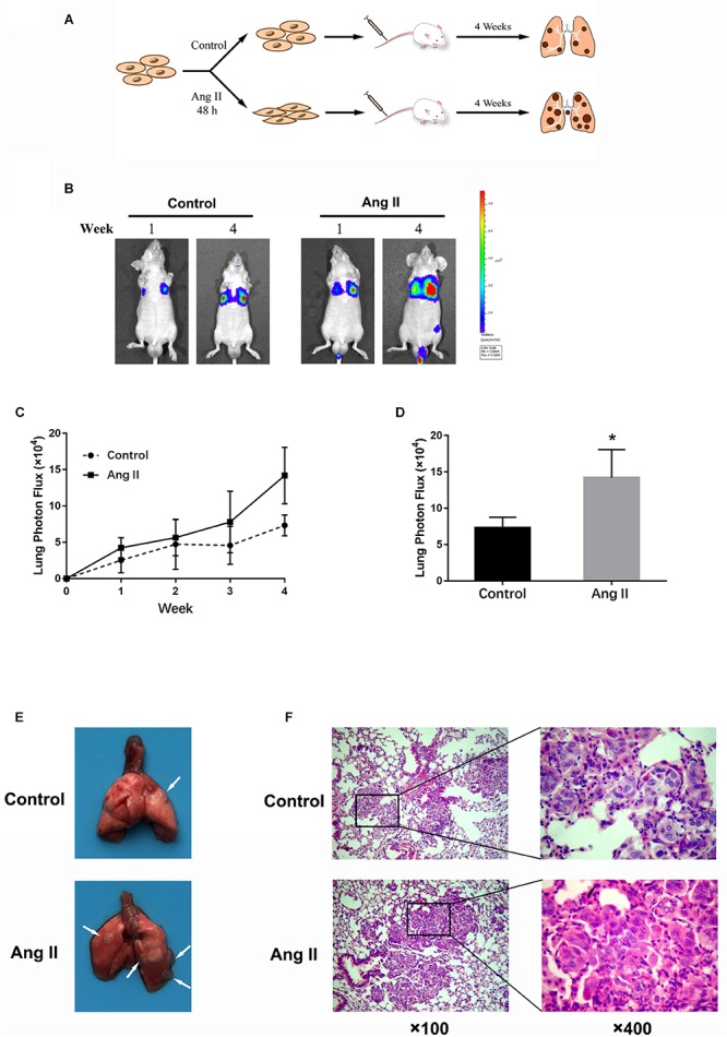
Ang II promotes NSCLC cell metastasis in vivo. (A) Schema of experimental protocol. Nude mice were intravenously injected with A549 cells pretreated with or without Ang II, as described in the Materials and Methods. After 4 weeks, mice were sacrificed, and lungs were imaged. (B) After injection at weeks 1 and 4, the mice were intraperitoneally injected with D-Luciferin for in vivo bioluminescence imaging. Tumor metastasis to lungs was shown. (C) Mean bioluminescence/time of lung metastasis in xenografted mice, graphed as normalized photon flux/time. (D) Mean bioluminescence at 4 weeks. (E) Representative images of lung metastatic nodules (arrows indicate tumor lesions). (F) Representative pictures of HE staining of the lung issue are shown (magnification, left × 100 and right × 400). *p < 0.05.
