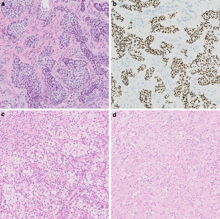
Fig. 1.

Case 1 (a index case) showed sheets of slightly polymorphic cells with clear to eosinophilic cytoplasm and positive nuclear staining for P63 (b). Case 3 (c) showed clear cells with intervening collagenic bundles and case 5 (d) was composed of strands and trabeculae of clear cells. HE, 20× (a, c, d). P63, clone 4A4, 1:3000, Immunologic, VWR International, Radnor, USA, 20x (b)
