
Figure 1.
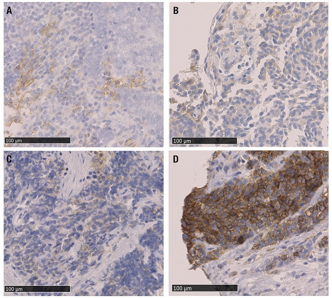
Microphotographs of representative examples of IHC PD-L1 expression in NEN G3 samples; different levels of staining (brown) are shown. (A) PD-L1 positive TC (score 1+) (gastrinoma, distant metastasis), (B) PD-L1 positive IC (score 1+) and PD-L1 negative TC (Merkel cell carcinoma lymph node metastasis), (C) PDL1 positive TC and IC, both score 1+ (lung, primary tumor sample), (D) PD-L1 positive TC (score 3+) (colon MiNEN, distant metastasis). PD-L1: programmed cell death ligand 1, IHC: immunohistochemistry, TC: tumor cells, IC: tumor-infiltrating immune cells, MiNEN: mixed neuroendocrine-nonneuroendocrine neoplasm.