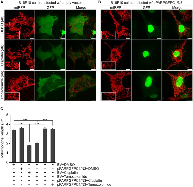
FIGURE 4.
Effect of PARPDN, cisplatin and temozolomide on mitochondrial fragmentation. B16F10 cells were co-transfected with either mtRFP and an empty vector (EV) (A,C) or mtRFP and pPARPGFPC1/N3 (B,C), treated as indicated in the figure for 4 h and the lengths of mitochondria were quantified. Panel (A,B) show representative reconstructions of confocal z-stacks while (C) displays the quantification of the mean length of mitochondria at 4-h post-treatments. Values are expressed as mean + SEM, N = 3, ∗p < 0.05, ∗∗p < 0.01, ∗∗∗p < 0.001. Scale bar: 10 μm.

