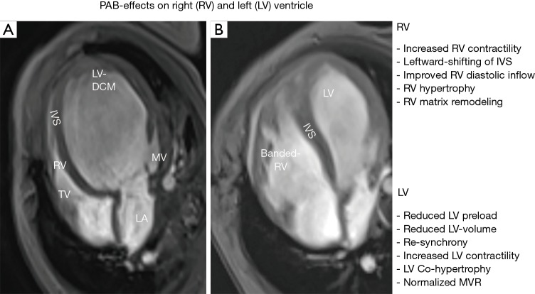
Figure 1.
Summarizes the pulmonary artery banding (PAB) effects on the right (RV) and left ventricle (LV). (A) Depicts the magnet resonance imaging (MRI) in four-chamber view of an infant with left ventricular dilated cardiomyopathy (LV-DCM); (B) shows functional regeneration of the LV based on the PAB induced ventriculo-ventricular interaction (VVI); the MRI was performed before the PAB induced RV hypertension was unloaded by transcatheter balloon dilation. IVS, interventricular septum; LA, left atrium; MV, mitral valve; TV, tricuspid valve.

