Figure 1.

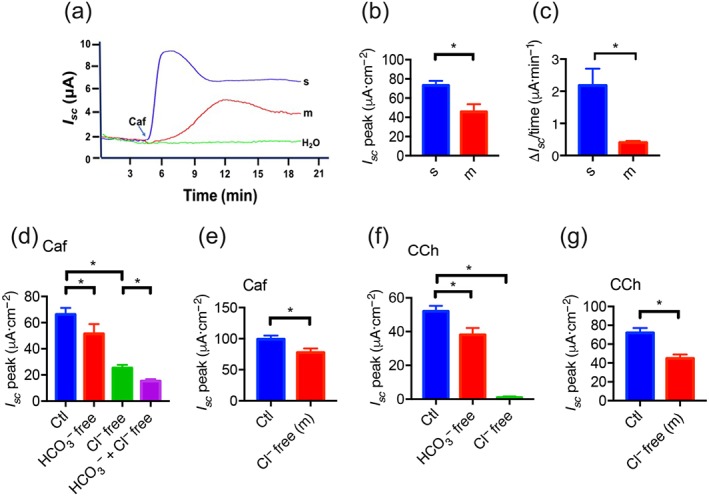
The effect of caffeine on murine duodenal epithelial anion secretion. (a) Representative time courses of caffeine (Caf; 10 mM)‐stimulated I sc or control (H2O) when added to the serosal (s) or mucosal (m) side of duodenal mucosal tissues. (b–c) Summary data of caffeine‐stimulated I sc peak and rising rate when added to the serosal or the mucosal side (n = 6). (d) Summary data of caffeine‐stimulated I sc peak after HCO3 − or Cl− omission from both mucosal and serosal sides. Ctl represents as the control with HCO3 − and Cl− in both mucosal and serosal sides. (e) Summary data of caffeine‐stimulated I sc peak after Cl− omission from the mucosal side (n = 7). (f) Summary data of carbachol (CCh)‐stimulated I sc peak after HCO3 − or Cl− omission from both mucosal and serosal sides. (g) Summary data of carbachol‐stimulated I sc peak after Cl− omission from the mucosal side (n = 6). * P < 0.05, significantly different from the corresponding control
