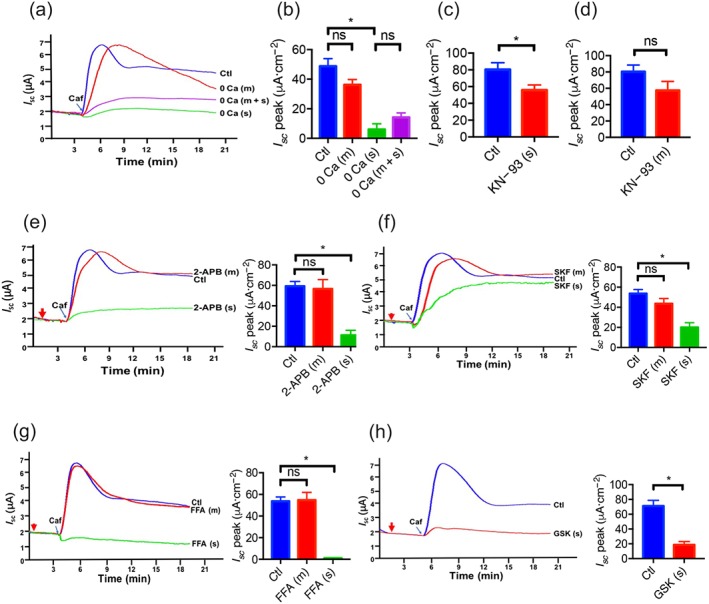
Figure 2.

The critical roles of Ca2+ signalling and Ca2+ channels in caffeine‐stimulated duodenal epithelial anion secretion. (a) Representative time courses of caffeine (Caf; 10 mM)‐stimulated I sc after extracellular Ca2+ omission (0 Ca) from mucosal (m), serosal (s) side, or both sides (m + s) of duodenal mucosal tissues. Ctl represents as the control in which 2‐mM extracellular Ca2+ was in both mucosal and serosal sides. (b) Summary data of caffeine‐stimulated I sc peak after Ca2+ omission from mucosal (m, n = 10) side, serosal (s, n = 11) side, or both sides (m + s, n = 8). (c–d) Summary data of caffeine‐stimulated I sc peak after serosal or mucosal addition of KN‐93 (10 μM, n = 6) in the presence of 2‐mM extracellular Ca2+. Ctl represents as the control without drug treatment. (e–g) Representative time courses and summary data of caffeine‐stimulated I sc peak after mucosal or serosal addition of 2‐aminoethoxydiphenyl borate (2‐APB, 50 μM, n = 7), SKF‐96365 (SKF, 30 μM, n = 7), or flufenamic acid (FFA, 100 μM, n = 9). The red arrows indicate the time of inhibitor addition. (h) Representative time courses and summary data of caffeine‐stimulated I sc peak after serosal addition of GSK‐7975A (GSK, 100 μM, n = 8). ns: no significant differences, * P < 0.05, significantly different from the corresponding control
