Figure 1.

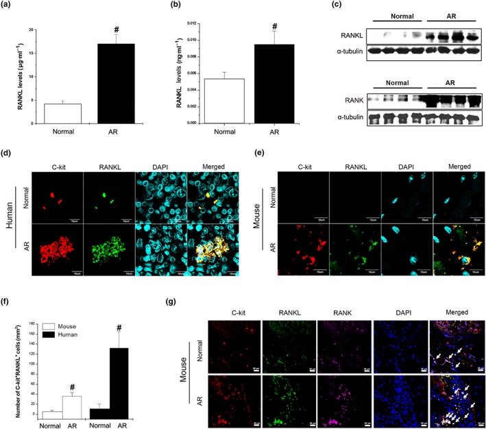
RANKL is up‐regulated in AR patients and localized on mast cells. (a) RANKL in serum (n = 40) and (b) homogenized nasal mucosal tissues (n = 20) of AR patients was analysed by elisa. # P < 0.05; significantly different from normal. (c) RANKL (upper panel) and RANK (lower panel) expression in the nasal mucosa of AR patients was determined by western blot analysis. (d) Immunostaining for RANKL (green) and staining for C‐kit (red) is shown in the AR patients (magnification, ×294). Mice were sensitized on Days 1, 5, and 14 by i.p. injections of 100 μg of OVA emulsified with 20 mg of aluminium hydroxide and then challenged with 1.5 mg of OVA. (e) Immunostaining for RANKL (green) and staining for C‐kit (red) is shown in nasal mucosal tissues from normal mice and AR mice (magnification, ×294). (f) C‐kit+RANKL+ cells were counted in AR patients and animal tissues by three individuals (n = 5 per group). Data shown are the means ± SEM. # P < 0.05; significantly different from normal. (g) Co‐localization of C‐kit (red), RANKL (green), and RANK (purple) in the nasal mucosa of AR animal (magnification, ×100). Arrows indicate C‐kit+RANKL+RANK+ cells
