Fig. 1.

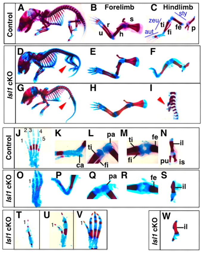
Hindlimb-specific defects in Hoxb6Cre; Islet1 conditional knockout mice. Skeletal preparations of newborn control (Hoxb6CreTg/+; Islet1flox/+; A-C,J-N) and mutant (Hoxb6CreTg/+; Islet1flox/-; D-I,O-W) mice. (A-C) Lateral views of the control mouse (A). In the forelimb (B), the scapula (s), humerus (h), radius (r) and ulna (u) are indicated, and in the hindlimb (C), pelvic girdle (p), femur (fe), tibia (ti) and fibula (fi) are indicated. Aut, autopod; sty, stylopod; zeu, zeugopod. (D-I) Lateral views of Isl1 cKO newborn with three digits in a leg (D-F) and a mutant lacking hindlimbs (G-I). In both cases, defects specific to the hindlimb (red arrowheads) were observed (D,G). Forelimbs formed normally (E,H) but only one zeugopodal bone formed in a mutant (F). In the mutant lacking hindlimbs, only the pelvic girdle is present (arrowhead, I) (J-N) Dorsal view of the hindlimb autopod (J), lateral views of the ankle (K) and knee (L) and dorsal views of the knee (M) and pelvic girdle (N) of a control mouse. Digits are numbered 1-5. The calcaneus (ca) in the ankle, and the patella (pa) in the knee are structures characteristic for hindlimbs. (O-S) Dorsal view of the hindlimb autopod with three digits (O). The calcaneus is missing in the ankle (P), but the patella is present in the knee (Q). The knee articulation in Isl1 cKO hindlimbs is similar to that in controls (R). The mutant pelvic girdle consists of an ilium (il) located anteriorly, whereas ischium (is) and pubis (pu) failed to develop. (T-W) Mutants with one digit (T), two digits (U) or four digits (V) were also obtained. In all cases the most anterior digit 1 was present and the most posterior digit (digit 5) was lost (O,T-V). In case of hindlimb aplasia, a pelvic girdle with a morphology similar to the other phenotypic groups formed (W).
