Fig. 7.

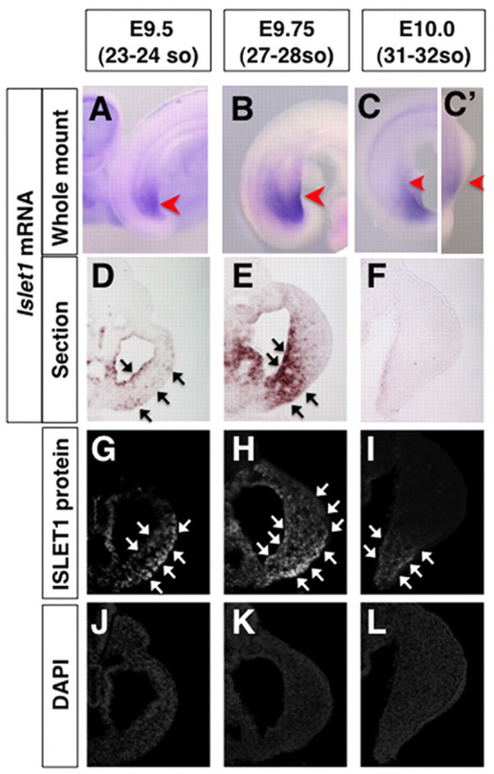
Spatial distribution of Islet1 mRNA and ISLET1 protein in hindlimb buds. (A-C′) RNA in situ hybridization showing the expression of Islet1 in the hindlimb field and bud. Red arrowheads indicate the approximate positions of sections shown in panels D-L. Dorsal-lateral views of the E9.5 (A) and E9.75 (B) embryos show the expression of Islet1 in the posterior hindlimb field. Lateral (C) and dorsal (C′) views of the E10.0 embryo show absence of Islet1 expression in hindlimb buds. (D-L) Islet1 section RNA in situ hybridization (D-F) and ISLET1 protein immunostaining (G-I) using adjacent sections, and DAPI analysis (J-L) of the sections shown in panels G-I. Immunoreactive ISLET1 proteins (G,H) were detected in more cells than were Islet1 transcripts (D,E) at E9.5 and E9.75. Islet1 mRNA was barely detectable in hindlimb buds at E10.0 (F), whereas ISLET1 proteins remained in the ventral part of the posterior hindlimb bud (I). Black and white arrows indicate Islet1 and ISLET1 positive areas, respectively.
