
Figure 3.
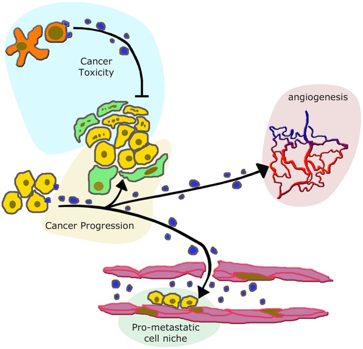
Extracellular vesicles (EVs) from immune and cancer cells induce different effects on cancer and peripheral structures. EVs of variable size, released from lymphocytes activated by dendrites (red cytoplasm over light blue background distributed at the top of the figure) can induce cell toxicity affecting both adjacent cells (here fibroblasts and mesenchymal stromal cells (MSCs) labelled by several points, distributed over a green cytoplasm) and cancer cells (points over yellow background) 18, 19, 20. In contrast, EVs released from unaffected cancer cells reinforce the adjacent fibroblast (green) and cancer (yellow) cells, distributed over the central golden background. This effect of EVs induces cancer progression 49, 50, 51, 52, 53, 54, 55. Moreover, the EVs of cancer cell origin move to the proximity of neighbouring vessels, thus inducing stimulation of angiogenesis (red of the vessel system over the pink background to the right) 46. EVs of cancer origin penetrate within vessels (bottom, light green background), where they induce the intra‐lumenal assembly of pre‐metastatic cell niches 45, 46, 47, destined to develop into metastases.