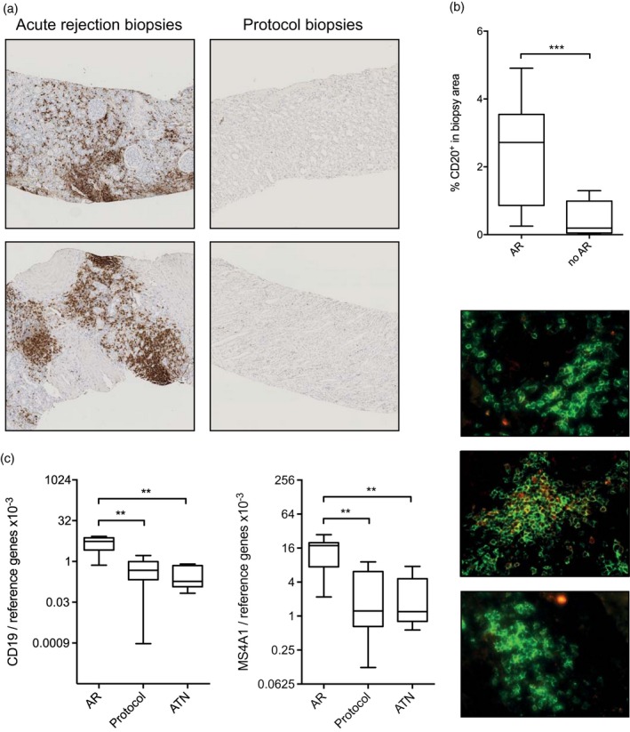
Figure 1.

Memory B cell infiltrates are present in grafts undergoing acute rejection (AR). (a) Representative examples of the presence of B cell infiltrates during AR and the absence of B cell infiltrates during stable graft function. (b) Quantification of CD20+ B cell infiltrates in biopsies from grafts undergoing AR and during stable graft function. AR: n = 16, no AR n = 7. (c) Gene expression levels for CD19 and MS4A1 are elevated in biopsies from grafts undergoing AR compared to biopsies during stable graft function and grafts showing acute tubular necrosis (ATN). AR: n = 7, protocol: n = 9, ATN: n = 7. (d) Representative examples of fluorescent stainings of CD20 (green) and IgD (red). Level of significance: *P < 0·05, **P < 0·01, ***P < 0·001.
