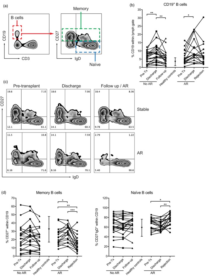
Figure 2.

The peripheral blood subset distribution is significantly altered at time of acute rejection (AR). (a) Flow cytometry gating strategy. (a) CD19+ B cells are increased at time of hospital discharge and normalize at time of rejection or follow‐up. (c) Representative examples of CD27 and immunoglobulin (Ig)D staining of a patient with stable graft function and a patient who underwent AR. (d) Memory B cell levels (defined by CD27 positivity) are increased at time of AR, coinciding with a decrease in naive B cells (defined by CD27 negativity and IgD positivity). Level of significance: *P < 0·05, **P < 0·01, ***P < 0·001.
