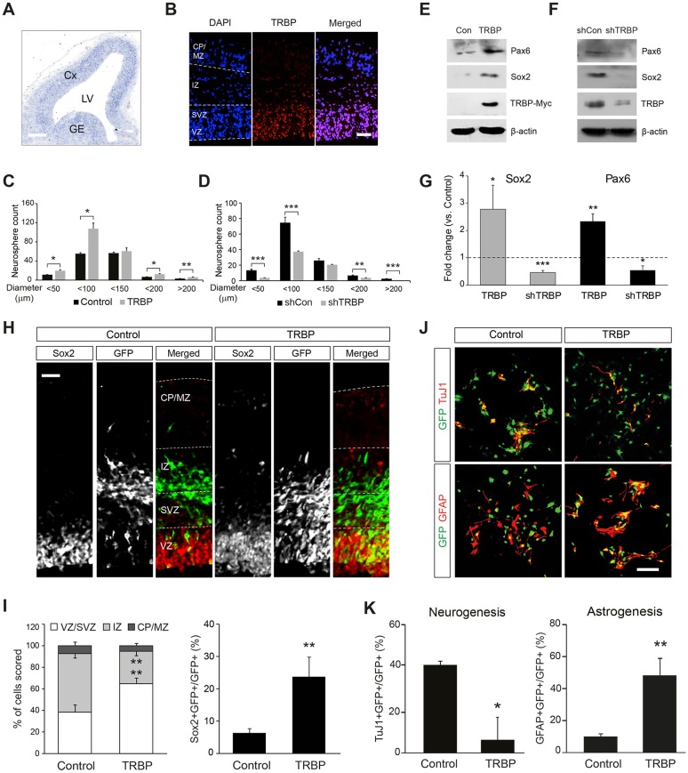
Fig. 1.
TRBP enhances embryonic neural stem cell properties. (A,B) Trbp gene expression pattern in the E13.5 mouse forebrain was determined by (A) in situ hybridization and (B) immunofluorescence after antigen retrieval using proteinase K digestion. (C,D) Neurosphere assay using mouse E14.5 primary neural progenitors transduced with a retroviral vector expressing (C) TRBP or (D) shRNA specific to TRBP (shTRBP). (E-G) Western blot analysis for Pax6 and Sox2 proteins of neural progenitor cell lysates infected with (E) TRBP-expressing or (F) shTRBP-expressing retrovirus. Quantification is shown in G. (H,I) Double immunolabeling of E15.5 brain sections electroporated in utero with TRBP-expressing plasmid at E13.5 using anti-GFP (reporter, green) and Sox2 (red) antibodies (H). Quantification is shown in I. (J,K) Representative immunostaining using anti-GFP (green) together with anti-βIII-tubulin (clone TuJ1; neuron, red) or GFAP (astrocyte, red) antibodies after in vitro differentiation of E14.5 neural progenitor cells. Quantification is shown in K. Scale bars: 200 µm in A; 50 µm in B,H,J. CP, cortical plate; Cx, cortex; GE, ganglionic eminence; IZ, intermediate zone; LV, lateral ventricles; MZ, marginal zone; SVZ, subventricular zone; VZ, ventricular zone. All error bars represent s.d. *P<0.05, **P<0.01, ***P<0.001, Student's t-test.

