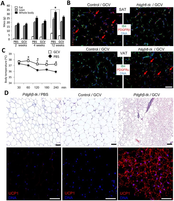
Fig. 4.
Depletion of the PDGFRβ+ lineage results in AT beiging. (A) Four-week-old Pdgfrb-tk mice (n=6/group) were injected intreperitoneally (i.p.) daily with 50 mg/kg or 100 mg/kg ganciclovir (GCV) or PBS control for 10 days. Data are fat and lean body mass measured using EchoMRI, along with whole body mass at the three time points post-treatment. (B) Paraffin wax-embedded sections of SAT from wild-type (control) and Pdgfrb-tk mice 1 month post-treatment with 100 mg/kg GCV were subjected to anti-PDGFRα or anti-PDGFRβ immunofluorescence (red) and endothelium-specific isolectin B4 (IB4) staining (green). Arrows indicate the comparatively high density of PDGFRβ+ or PDGFRα+ cells. (C) Body temperature of Pdgfrb-tk mice 1 month post GCV or PBS treatment measured over 240 min at 4°C. There is increased cold tolerance of PDGFRβ+ lineage-depleted mice. Data are mean±s.e.m. for multiple mice; *P<0.05 (Student's t-test). (D) Paraffin wax-embedded sections of SAT from Pdgfrb-tk or C57BL/6 (control) mice 1 month post-treatment with PBS (control) or 100 mg/kg GCV were subjected to Hematoxylin and Eosin staining (top) and anti-UCP1 immunofluorescence (red, bottom). Scale bars: 50 µm. Nuclei are blue. Experiments were repeated at least three times with similar results.

