Fig. 5.
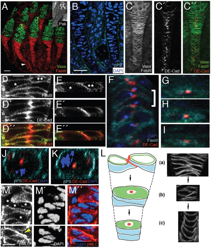
Cell intercalation in the pupal ovary forms a basal stalk with a novel radial cell polarity. (A-M″) Pupal ovaries 40 h after puparium formation (APF). Anterior end of each ovary is at the top. (A) Wild-type pupal ovary stained with anti-Vasa (green) to mark germ cells and with anti-FasIII (red) to mark basal stalk cells. Basal stalks are two cells wide at the anterior end of stalk where they contact the germ cells, but a single cell wide for most of their length, with bottle-shaped cells in the middle and disk-shaped cells at the posterior end (arrowhead). Inset shows anti-Pak staining of pupal ovary. Pak is expressed in basal stalk and germline cells. (B) Wild-type basal stalks stained with anti-βPS integrin to show that basal stalks are surrounded by basement membrane. (C-C″) Wild-type basal stalks stained with anti-DE-Cadherin (red) to mark adherens junctions and with anti-Vasa and anti-FasIII to reveal cells (green). There are adherens junctions where cells contact each other in the two-cell-wide proportion of the basal stalk and adherens junctions between cells in the single-cell-wide proportion. (D-D″) High-magnification view of adherens junction between two cells (asterisks) in two-cell-wide proportion of wild-type basal stalk. (E-E″) Cross-sectional view of same cells as in D-D″. (F) Bottle-shaped cells in wild-type basal stalk stained with DAPI (blue) to mark nuclei and anti-FasIII (cyan) and anti-DE-Cadherin (red), showing ‘yin-yang’ arrangement, in which narrow end of one cell is in contact with wide end of its neighbors. (G-I) Bottle-shaped cells marked by the bracket in F, viewed in cross-section. Cells contain a dot of DE-cadherin staining surrounded by FasIII staining. (J,K) Neighboring disk-shape cells from a basal stalk showing DE-Cadherin at center of disk (red) and basal marker βPS integrin (cyan) circumferentially distributed. Nuclei are stained with DAPI (blue). The distribution of polarity markers in G-K indicates a radial cell polarity. (L) Schematic showing the three types of cell organization in the basal stalk. Red, DE-Cadherin; green, FasIII; blue, βPS integrin. (a) At the anterior end the stalk is two cells wide, with adherens junction at point of contact. (b) In the middle of stalk, cells are bottle-shaped in side view and linked by spot adherens junctions. (c) At the posterior end of stalk cells are disk-shaped. Note the radial cell polarity in (b) and (c). We propose that the three types of organization are stages in the intercalation process (arrows). (M-M″) Wild-type basal stalk stained with anti-pMLC and DAPI. Some cells show no pMLC staining (asterisks), whereas others show high levels. Cell marked with arrowhead in K shows high level of pMLC in constricted region. Scale bars: 10 µm in A,B for A-C″; 2 µm in D for D-K; 4 µm in M for M-M″.

