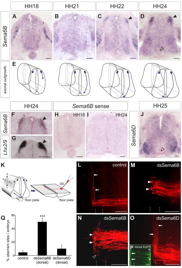
Fig. 1.
Sema6b expression in dI1 commissural neurons peaks at HH24 and is required for post-crossing commissural axon guidance. (A-E) Expression of Sema6b mRNA in transverse sections of the chicken lumbar spinal cord at the indicated developmental stages (A-D) with schematic representations of the corresponding growth of dI1 axons (E). Sema6b is first detected in dI1 neurons at HH22, when their axons have reached the ipsilateral floorplate border (C, arrowhead). Expression of Sema6b in dI1 neurons is strongest at HH24 (D, arrowheads). (F,G) Probing adjacent sections with Lhx2/9 confirmed that Sema6b is expressed in dI1 neurons (arrowheads). (H,I) No staining is seen with a Sema6b sense probe. (J) Diffuse expression of Sema6d at HH25, with higher levels in motoneurons. Neither Sema6b nor Sema6d is expressed in the floorplate (D,J, open arrowheads). (K) Schematic of an open-book preparation and application of DiI for axonal tracing (red). D, dorsal; V, ventral; R, rostral; C, caudal. (L) Commissural axons cross the midline and extend along the contralateral floorplate border (arrows) in untreated control embryos. (M,N) After silencing Sema6B by injection and electroporation of dsRNA (dsSema6b), axons stall at the contralateral floorplate border (closed arrowheads) or even turn caudally (open arrowhead). (O) Silencing Sema6D by injection and electroporation of dsRNA (dsSema6d) did not affect axon guidance (arrows). (P) Expression of a co-electroporated EGFP reporter confirmed the exclusively dorsal targeting of dsRNA (asterisks). Arrows mark axons of contralaterally projecting commissural neurons. The floorplate is indicated by dashed lines. (Q) Quantification of DiI injection sites with aberrant axonal pathfinding. ***P<0.001; error bars indicate s.e.m. Scale bars: 50 µm in A-D,F-J; 100 µm in L-P.

