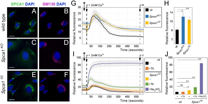
Fig. 3.
Effects of the 1D mutation on the localization and function of SPCA1. (A-F) Immunohistochemistry with SPCA1 (A,C,E, green) and GM130 (GOLGA2) (B,D,F, magenta) antibodies in wild-type (A,B), Spca1KO (C,D) and Spca11D (E,F) MEFs. Scale bars: 20 µm. (G-J) Calcium clearance assays in wild-type (black, n=9), Spca1KO (blue, n=6) and Spca11D (yellow, n=9) MEFs in the absence (G,H) or presence (I,J) of drug inhibitors. Drug treatments were as follows: wild-type MEFs treated with 100 nM thapsigargin (Th; SERCA inhibitor, orange, n=8), Spca11D MEFs treated with 100 nM thapsigargin (green, n=7) and Spca11D MEFs treated with sodium orthovanadate (violet, n=4). At time 0, cells were spiked with 2 mM Ca2+ (black dashed lines). Resting cytosolic calcium levels 10 min after introduction of calcium (gray dashed lines) are quantified in H and J. Asterisks in G and I indicate measurements in resting conditions before the addition of Ca2+. Error bars indicate s.e.m. ns, not significant; *P<0.05, **P<0.01.

