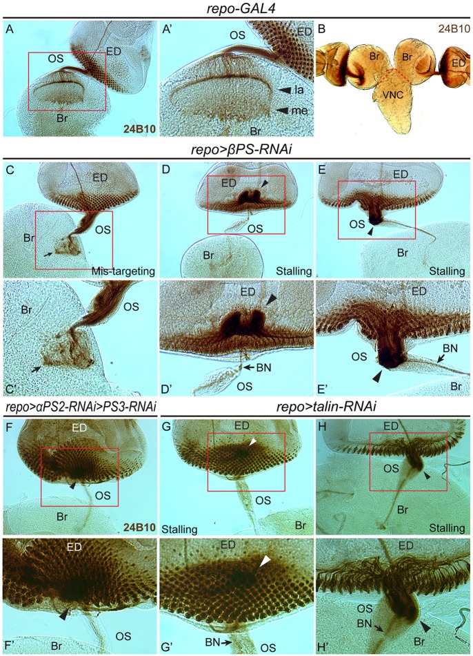
Fig. 4.
Photoreceptor axon migration defects observed with focal adhesion loss in glia. Photoreceptors were immunolabeled with anti-Chaoptin antibody (mAb 24B10). (A-B) In control larvae, photoreceptor axons exited the ED, passed through the OS and terminated in the lamina (la) and medulla (me) (A′) in the brain lobe (Br). VNC, ventral nerve cord. (C-E′) In repo>βPS-RNAi larvae, the photoreceptor axons exhibited a disorganized pattern in the brain lobe (C,C′, arrow), failed to exit the ED (D,D′, arrowheads) or stalled in the OS (E,E′, arrowheads). BN, Bolwig's nerve. (F-H′) Axon stalling phenotypes were also observed in wandering third instar larvae of repo>αPS2-RNAi>αPS3-RNAi and repo>talin-RNAi. Boxed regions are magnified in the respective prime panels.

