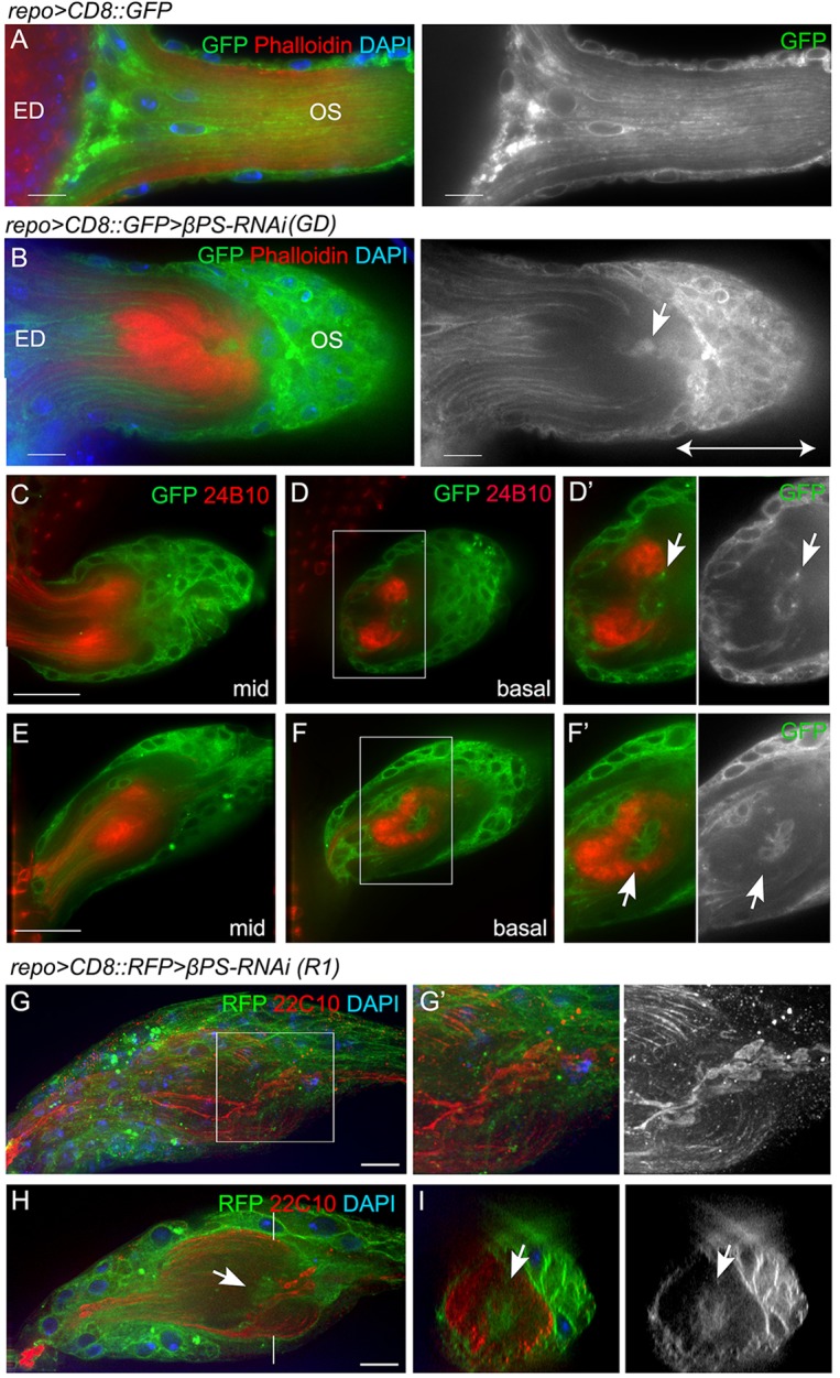
Fig. 5.

Axon stalling within the OS. (A,B) Alexa 568-phalloidin (red) was used to label F-actin in control (A) and repo>βPS-RNAi (B) ED and OS. Glial membranes were labeled with CD8::GFP (green) and nuclei are marked with DAPI (blue). A glial cap (double-ended arrow) formed proximal to the stalling region, with glia (arrow) present in the stalling region. (C-F′) Glia are associated with stalled axonal terminals. Single optical sections from the middle of the z-stack (mid) or from the basal level are shown. Digital magnifications of boxed regions are displayed to the right in grayscale to illustrate the glial membranes. Arrows point to the glia associated with the stalled axon terminals. (G-I) Immunolabeling for Futsch (mAb 22C10, red) marks microtubules in the photoreceptor axons in repo>βPS-RNAi-treated OSs. A projection of the entire stack (G) with the area of axon stalling (G′) are shown. A single optical section from the middle of the stack (H) and the orthogonal sections (I) highlight the stalling region and the glia within it (arrows). All panels except G are single 0.2 μm z-sections. Scale bars: 10 μm.
