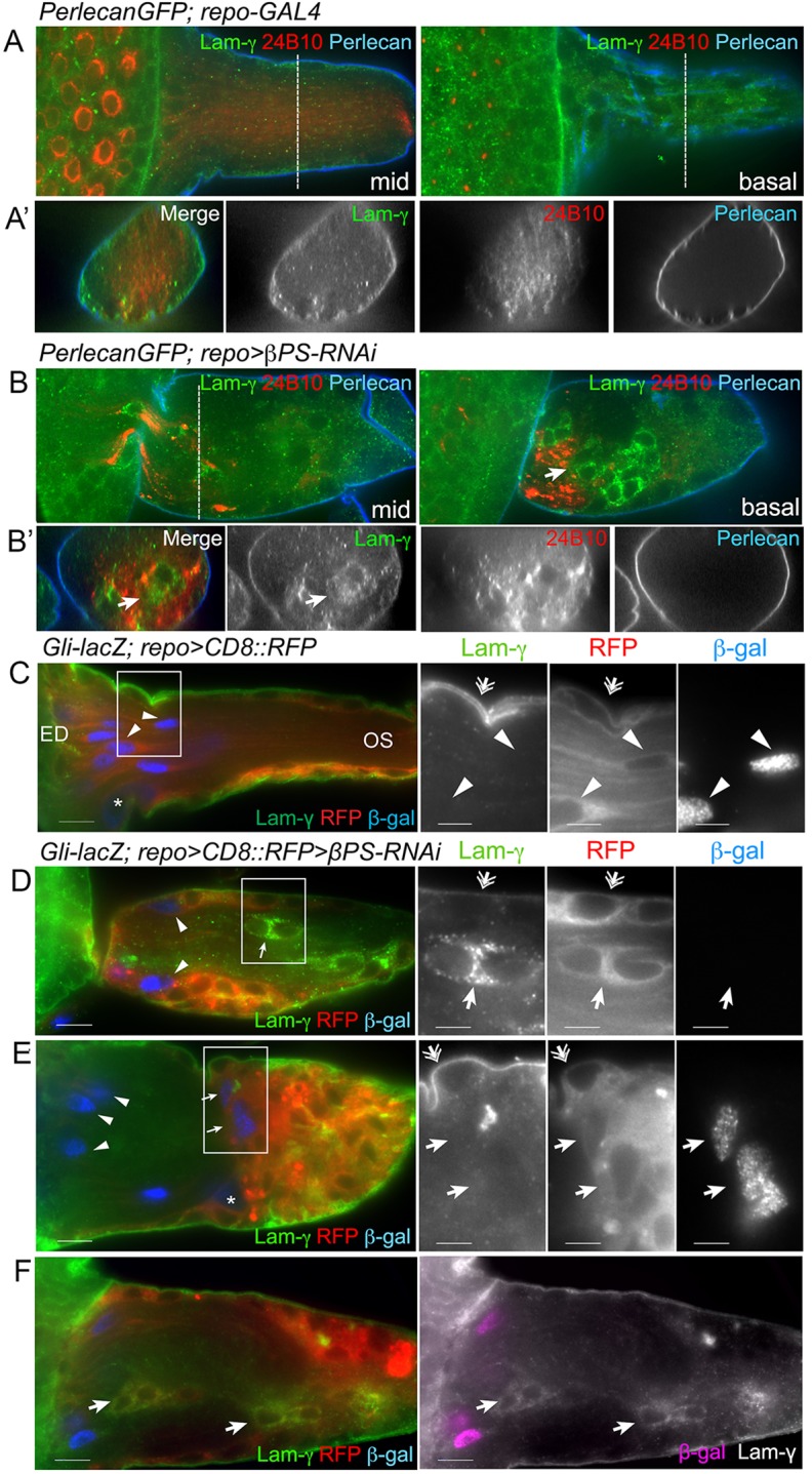
Fig. 7.

Ectopic glia express PG markers. (A-B′) LanB2 (Lam-γ, green) and Perlecan::GFP (blue) were used to label the ECM. Photoreceptor neurons were immunolabeled with anti-Chaoptin antibody (mAb 24B10). Two optical sections are shown: midway (mid) and basal. Transverse sections (dashed lines) are shown in the lower panels. In controls (A), both ECM markers were predominantly found in the outermost neural lamella, with only diffuse LanB2 labeling inside the glia. In repo>βPS-RNAi, Perlecan::GFP was detected in the neural lamella but glia expressing LanB2 were seen inside the OS associated with stalled axons (arrows). (C-F) LanB2 (green) and Gli-lacZ (β-gal, blue) marked PG and differentiated WG in control (C) and repo>βPS-RNAi (D-F) OS. Glial membranes were labeled with repo>CD8::RFP (red). Digital magnifications of boxed regions (C-E) are displayed to the right. In controls (C) LanB2 was predominant in the outer ECM and PG (double arrows), but not in the WG, which were distinguished by long membrane processes and Gli-lacZ expression (arrowheads). In repo>βPS-RNAi (D-F), LanB2 was detected in the ECM (double arrows) and surface PG. LanB2-positive glia (D,F, arrows) were observed in the OS core along with WG (arrowheads). In some OSs, Gli-lacZ-positive WG were observed in their normal position (E, arrowheads) and in the glial cap proximal to the axonal stalling regions (E, arrows). The majority of OSs had LanB2-positive Gli-lacZ-negative glia within the stalling region and in the glial cap (F, arrows). All panels are single 0.2 μm z-sections. Scale bars: 10 μm (left panel); 5 μm (right panels in C-E).
