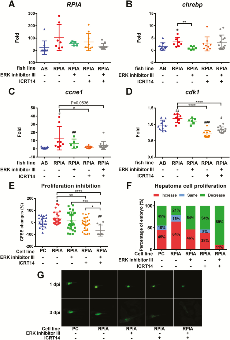
Figure 6.
ERK inhibition diminished lipogenesis while β-Catenin inhibition reverted cellular proliferation in zebrafish. (A–D) QPCR was used to measure the relative mRNA fold induction of RPIA, lipogenic factor (chrebp) and cell cycle/proliferation markers (ccne1 and cdk1) in two independent lines of Tg (fabp10a: RPIA) compared with the control fish (mC) after ERK inhibitor III and β-catenin inhibitor (ICRT14) treatment by oral feeding. (E) PLC5 with RPIA-overexpressed cells proliferation ability was examined by xenotransplantation assay. The CFSE‐labeled cells were injected to 2-day-old embryos. PLC5 was pretreated with ERK inhibitor and β-catenin inhibitor or mixture of these two inhibitors for 24 h before injection. The cell proliferation was analyzed between 1 and 3 dpi. RPIA-overexpressed PLC5 (RPIA) has higher proliferation rate than the empty vector control (pcDNA3; PC). β-catenin inhibition (ICRT14) significantly reduced RPIA-expressed cell proliferation ability. Statistical analysis was performed by unpaired two-tailed t-tests with Welch’s correction. P < 0.05 was considered to be statistically significant. Hashtag (#) represented the comparison between RPIA-transgenic lines and AB; Asterisk (*) represented the comparison between RPIA Tg lines with and without inhibitor treatment. #0.01 < P ≤ 0.05; ###P ≤ 0.001; **0.001 < P ≤ 0.01; ****P ≤ 0.0001. (F) The percentage of embryos with increased, the same or decreased CFSE‐labeled cells compared between 3 and 1 dpi. β-catenin inhibition decreased the proportion of fish with RPIA-induced proliferative cells. Combination of β-catenin and ERK inhibitors revealed synergistic inhibition on RPIA-induced cell proliferation. (G) Representative fluorescence images of xenotransplanted zebrafish at 1 and 3 dpi for each treatment.

