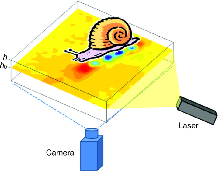
Fig. 7.
Schematic of the experimental set-up. As the animal crawled on the gelatin substrate (thickness h=8 mm), the deformation of the gel was measured by tracing the displacement of the embedded marker beads, which were illuminated by the planar laser sheet (h0=7.9 mm). Time-lapse images (15 frames s–1) of the illuminated laser plane were recorded. The deformation vector at each point was calculated by applying correlation techniques.

