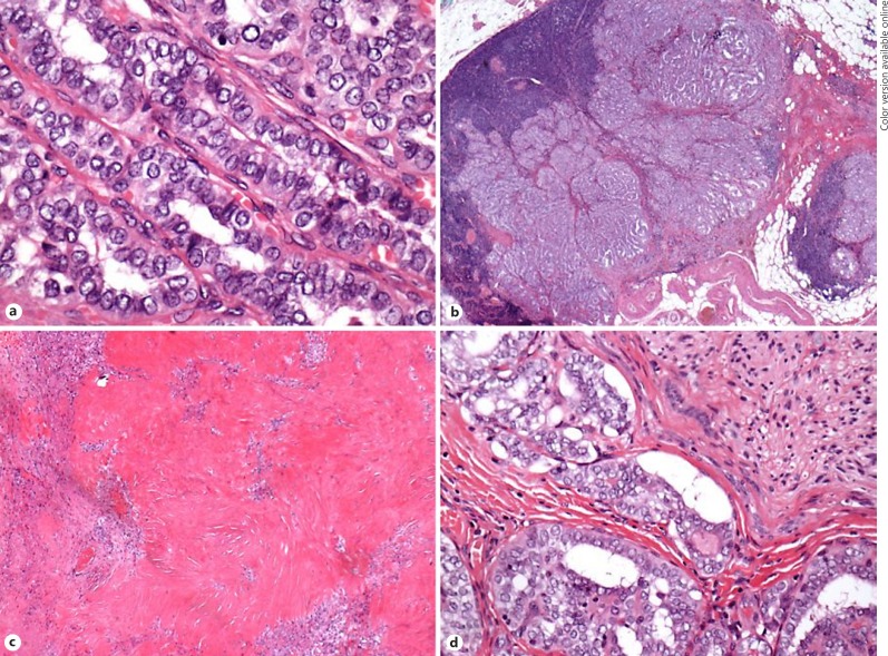
Fig. 4.
Tumour resection specimen (a; ×40 magnification): features of a well-differentiated papillary carcinoma are evident. No features of a poorly differentiated tumour. b Direct spread of tumour into a lymph node (×2 magnification). c Hyalinisation and fibrosis around tumour islands suggesting tumour regression secondary to TKI therapy (×10 magnification). d Perineural invasion (×40 magnification).

