Figure 1.
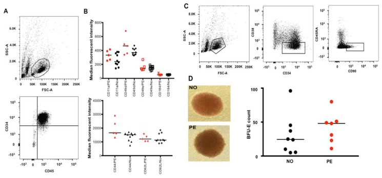
Flow cytometry analysis and isolation of UCB HSPCs and HSCs as well as assessment of surface adhesion molecules (SAMs) and in vitro erythroid differentiation capacity of the cells isolated from PE vs. normotensive (NO) pregnancies. (A) Flow cytometry analysis showing the UCB HSPC population gated based on size and granularity (FSC-A and SSC-A) and CD34+ CD45+ expression. (B) Demonstrating the median fluorescent intensity (MFI) for various SAMs in red (PE, n = 5) and black (NO, n = 10); despite large differences in some MFI values, the differences were not statistically significant. (C) Flow cytometry analysis of the HSC population from UCB samples; the population was gated (from left to right) based on size and granularity followed by CD34+, CD38lo, and CD45RA−, CD90+ expression. As previously reported by others, the CD34+ CD38lo population was very small in the majority of our samples. This specific individual sample with a large CD34+ CD38lo population was particularly chosen for specifically visualizing a clearly distinct CD34+ CD38lo CD45RA− and CD90+ population in the figure. (D) Example of BFU-Es in culture (10× magnification) from normotensive (n = 8) and PE (n = 7) samples after the UCB CD34+ cells were cultured for 14 days. No significant difference was observed BFU-E count comparison between PE and normotensive groups.

