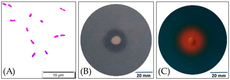
Figure 1.
Image of Gram-negative rod-shaped cells of the S17 isolate under light microscopy (A). Clear halo-zone of phosphate solubilization around the S17 isolate colony on a Pikovskaya (PVK) modified medium after 48 h of inoculation at 20 °C (B). Orange halo-zone around the colony of S17 isolate after 24 h of inoculation at 20 °C on the chrome azurol S blue medium (C).

