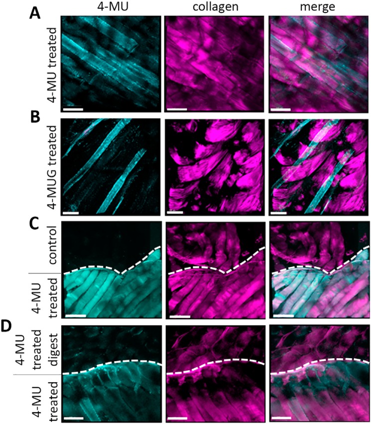
Figure 6.
4-MUG fluorescence can be detected in tissues via 2-photon imaging. A and B, representative 2-photon images of muscle tissue from 4-MU–treated mice (A) and 4-MUG–treated mice (B) show a specific signal in the 4-MU channel at a wavelength of 810 nm. C, representative 2-photon images of muscle tissue from untreated control mice (upper part) and 4-MU treated mice (lower part). D, representative 2-photon images of muscle tissue from 4-MU treated mice, where the muscle tissue from one mouse (upper part) was hyaluronidase digested. Untreated muscle tissue from a 4-MU treated mouse (lower part) serves as control. Upper and lower parts are indicated via a dashed line drawn in the picture. In each of those tissues 4-MU has a specific distribution as shown at 810 nm wavelength. Collagen was visualized at 920 nm, the 4-MU and collagen channel were merged for better structural orientation in the tissue. Scale bar = 100 μm.

