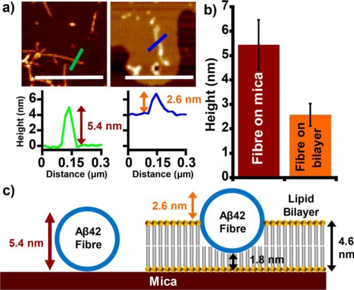
Figure 3.

Aβ fibers embed into the lipid bilayer, displacing the upper leaflet. a, AFM topographical images show Aβ42 fibers on mica (left) and on the surface of a lipid bilayer (right). Scale bar, 1 μm. Height scale range, 12 nm. Each image is accompanied by a height cross-section. b, mean Aβ42 fiber height recorded both on mica and above the bilayer surface. c, a scaled schematic showing how Aβ fibers displace the upper leaflet of the membrane as fibers laterally embed into the bilayer. Error bars, S.E.
