Figure 1.
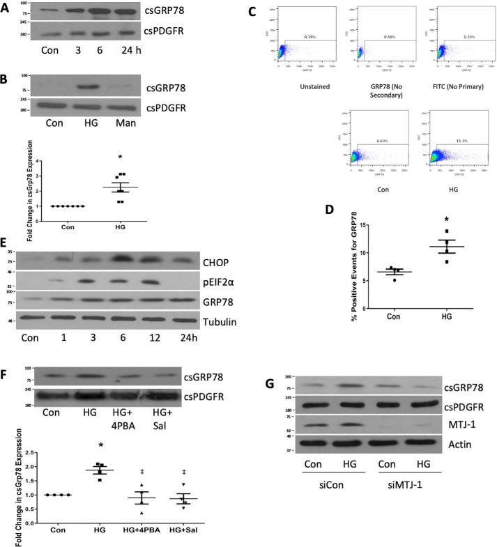
High glucose promotes the expression of GRP78 at the cell surface. A, MCs were treated with HG for the indicated times, and then csGRP78 was assessed by a biotinylation assay as described under “Experimental procedures.” PDGFR was used as a loading control (Con). B, csGRP78 was assessed by biotinylation after HG or mannitol (Man, 24 h, n = 7). C and D, expression of csGRP78 after HG for 24 h was confirmed by flow cytometry (n = 4). E, markers of ER stress were assessed by immunoblotting after HG treatment for the indicated times (n = 3) CHOP, CCAAT-enhancer-binding protein homologous protein. F, the effects of ER stress inhibitors 4-PBA (2.5 μm) or salubrinal (Sal, 30 μm) on HG-induced csGRP78 expression were assessed by biotinylation (n = 5). G, the effect of MTJ-1 down-regulation using siRNA on HG-induced csGRP78 expression was assessed by biotinylation (n = 3). MTJ-1 down-regulation was assessed by immunoblotting of whole-cell lysate. *, p < 0.05 HG versus control; ‡, p < 0.05 versus HG.

