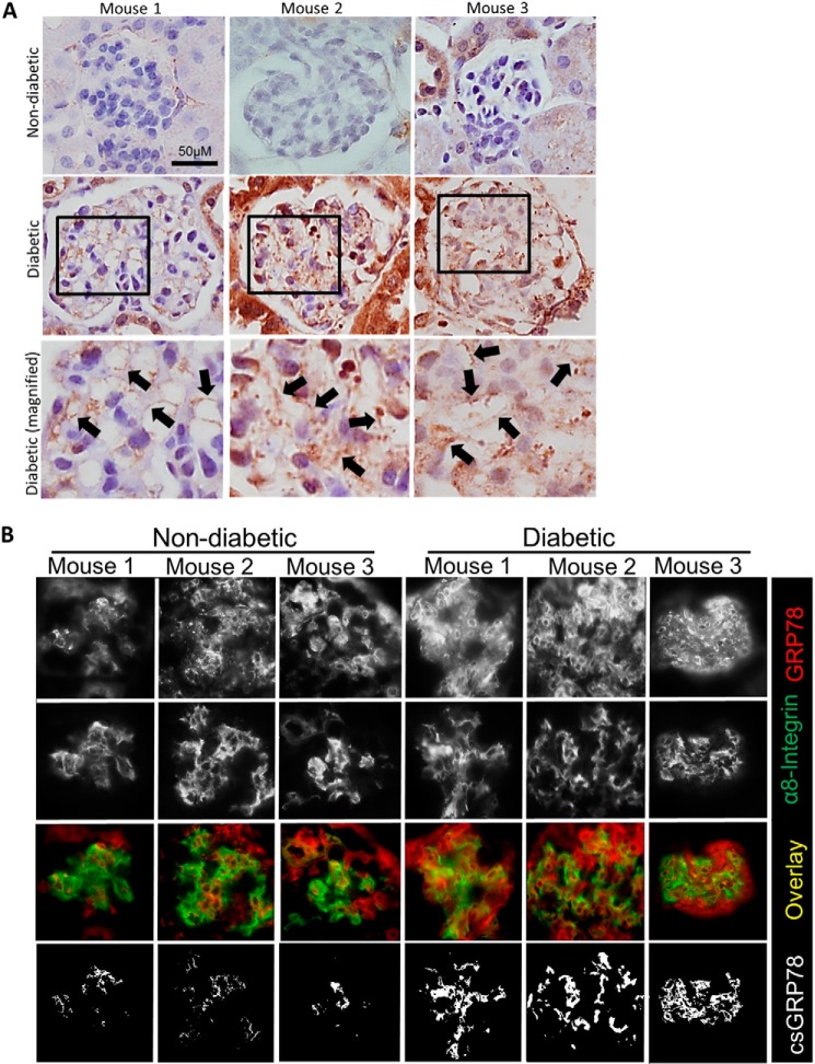
Figure 6.
Plasma membrane GRP78 is seen in glomeruli of type I diabetic CD-1 mice. Kidneys were harvested from vehicle- or streptozotocin-treated CD-1 mice after 12 weeks of diabetes (n = 3/group). A, paraffin-embedded sections were stained for GRP78. Black arrows indicate areas in which GRP78 appears to be localized to the plasma membrane. B, OCT sections were stained for GRP78 (red) or the plasma membrane marker α8-integrin (green). Areas of colocalization between GRP78 and the plasma membrane are indicated by white in the overlay and mask images.

