Figure 11.
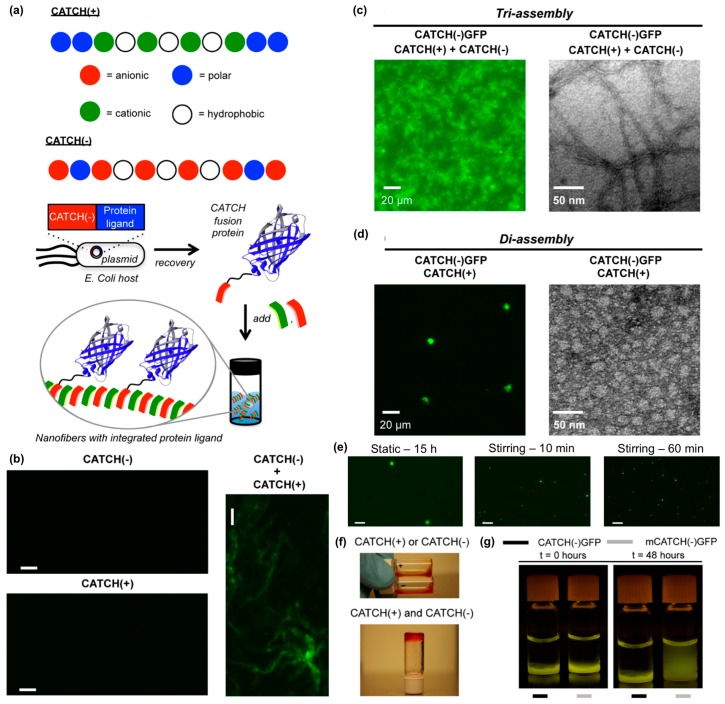
Co-assembling peptide tags for protein immobilization within nanofibrillar hydrogels. (a) Schematic representation of the Co-Assembly Tags based on CHarge complementarity (CATCH) nanofiber system. (b) Fluorescence photomicrographs of solutions containing Thioflavin T (ThT) and CATCH(−) only, CATCH(+) only, or an equimolar mixture of CATCH peptides. Nanofibrous structure was only observed in mixtures containing both peptides. Fluorescence photomicrograph (left) and transmission electron micrograph (right) of (c) CATCH(−), CATCH(+), and CATCH(−)GFP tri-assemblies, or (d) CATCH(+) and CATCH(−)GFP di-assemblies. Triassemblies formed nanofibrous structures, while diassemblies formed micron-sized particles. (e) Fluorescence photomicrographs of di-assembly microparticles after 15 h of static incubation (left), 10 mins of stirring (middle), and 60 mins of stirring (right). A notably higher number of microparticles having a smaller diameter formed with stirring compared to static control group. (f) Digital still images of aqueous buffered solutions of CATCH(+) and CATCH(−) alone (top), and hydrogel formed after mixing at a 1:1 (v/v) ratio (bottom). * Red food coloring was added to samples for ease of viewing. (g) Blue-light trans-illuminated digital still images of hydrogels fabricated by mixing CATCH(−) and CATCH(+) with 0.75 µM CATCH(−)GFP or 0.75 µM mutant CATCH(−)GFP (“mCATCH(–)GFP”) before and after 48 hours of incubation in excess 1× phosphate-buffered saline (PBS). CATCH(–)GFP was retained within the hydrogel over time while mCATCH(–)GFP was released into the surrounding medium. Scale bar = 20 μm in (b) and (e). Adapted with permission from [114]. Copyright 2016, Springer US publishing.

