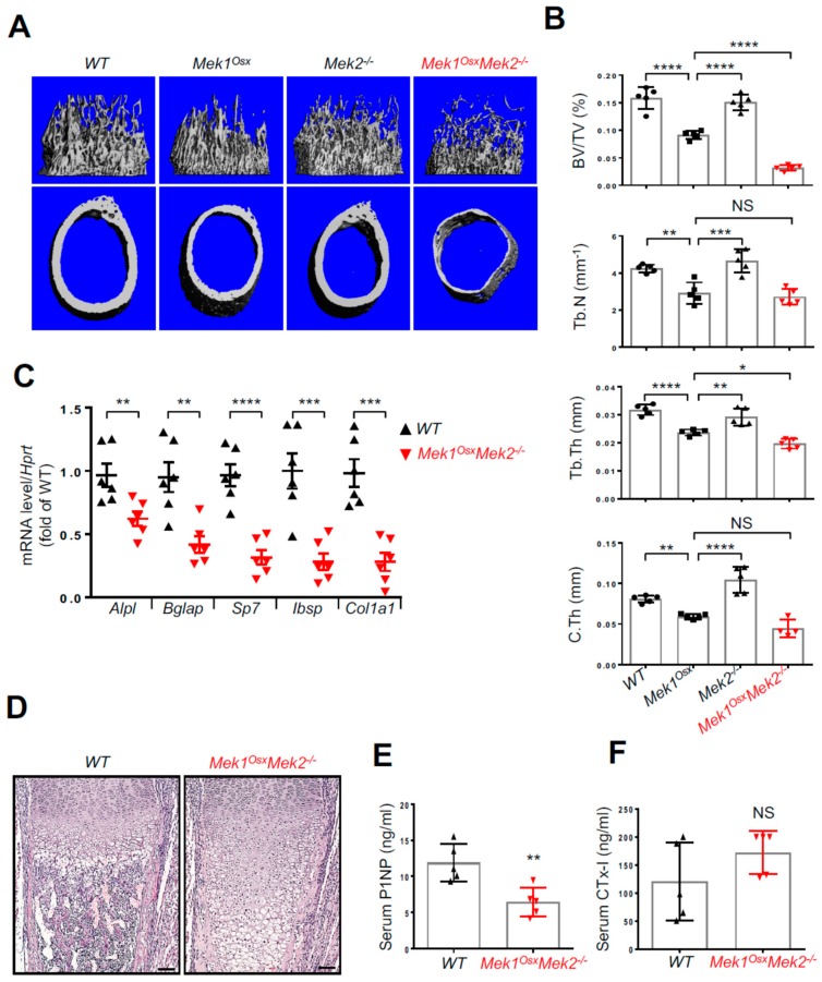
Figure 3.
Inactivation of ERK in osteoprogenitors causes severe osteopenia in long bones. (A,B) MicroCT analysis of three-week-old WT, Mek1Osx, Mek2−/−, and Mek1OsxMek2−/− femurs. Representative 3D-reconstruction images of trabecular (upper) and cortical bone (lower) are displayed (A). Quantification of trabecular bone mass and midshaft cortical bone thickness are displayed (B). Trabecular bone volume/total volume (BV/TV), trabecular thickness (Tb.Th), trabecular number per cubic millimeter (Tb.N), and cortical thickness (C.Th). (n = 4~5). (C) Total RNAs were isolated from three-week-old WT and Mek1OsxMek2−/− tibias, and mRNA levels of osteoblast differentiation genes were measured by RT-PCR. (n = 6). (D) H&E-stained longitudinal sections of three-week-old WT and Mek1OsxMek2−/− femurs. Scale bar, 1 mm. (E,F) Serum levels of P1NP and CTx-I were measured by ELISA. (n = 5). Values represent the mean ± SD.; NS, not significant, * p < 0.05, ** p < 0.01, *** p < 0.001, and **** p < 0.0001 by the one-way ANOVA test (B) or an unpaired two-tailed Student’s t-test (C,E,F).

