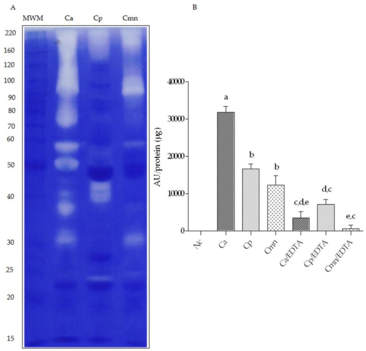
Figure 4.
Gelatin zymography in 10% polyacrylamide gels and proteolytic activity using casein substrate. (A) Ten micrograms of venom protein from C. aquilus (Ca), Crotalus polystictus (Cp) and C. molossus nigrescens (Cmn) were incubated after electrophoresis for 2.5 h at 37 °C. MWM—molecular weight markers (kDa). (B) Proteolytic activity of 100 μg of venom samples in the presence/absence of 50 mM EDTA incubated with casein as a substrate for 2.5 h at 37 °C. Negative control (Nc). Small letters (a,b,c,d,e) show statistical difference (Tukey p < 0.05). Variability in proteases bands is marked with red boxes. Small letters show significant differences (Tukey p < 0.05) between samples, either with or without ethylendiaminetetraacetic acid (EDTA).

