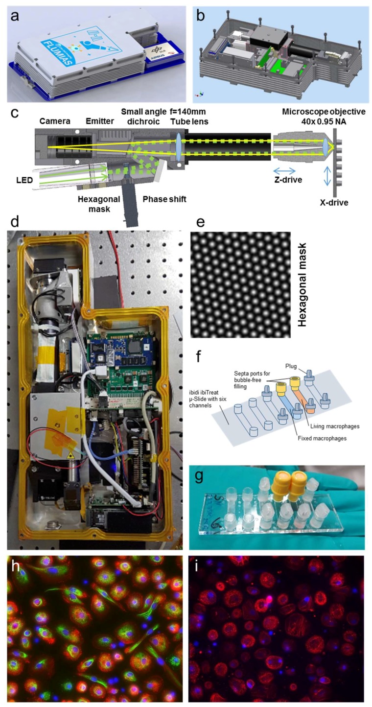
Figure 1.
The FLUMIAS-DEA microscope. (a) CAD-image of the microscope attached to the Tango Lab carrier card. (b) CAD-image with the double sealed cover removed. (c) Optical beam path of the microscope. The excitation light coming from the LED is structured by passing the hexagonal mask and moved by the phase shift motor. A quad-band small angle dichroic is reflecting the excitation light towards the sample passing the tube lens and focused by the Nikon 40×, 0.95 NA air corrected objective. The objective is moved by a piezo to acquire z-stacks. The ibiTreat µ-Slide VI 1.9 (custom made by ibidi, Martinsried, Germany, channel volume app. 120 µL, referred to as ibidi µ-Slide) integrated into the sample holder can be moved on one axis to address two of the six ibidi µ-Slide channels. The fluorescence light passing the dichroic mirror is filtered by a quad band emitter and is detected by the camera. (d) Photograph of the microscope. (e) Intensity distribution of the structured illumination patterned excitation. (f) Scheme of cell culture vessel with living and fixed macrophages. (g) Photograph of ibidi µ-Slide with living and fixed macrophages. (h) Three channel overlay of maximum projected SIM-images of the fixed sample; size is 370 × 370 µm. Staining and imaging parameters of the fixed cells can be found in Table 2. (i) Two channel overlay of maximum projected SIM-images of the living macrophage sample; size is 370 × 370 µm. Staining and imaging parameters of the living cells can be found in Table 2.

