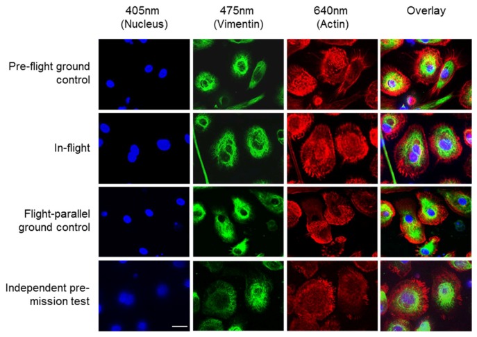
Figure 2.
Fluorescence microscopy images of fixed primary human macrophages exposed to microgravity on the ISS. Cells were stained for nuclei (DAPI, blue), vimentin (anti-vimentin-Alexa Fluor 488; green) and actin (SiR-actin, red). Images were acquired on ground before upload to the ISS (Pre-flight ground control), and after 4 days of exposure to microgravity on the ISS (In-flight). Also shown are pictures of cells that were cultured and imaged parallel to the flown cells (Flight-parallel ground control) and images from a pre-test of the FLUMIAS-DEA microscope (Independent pre-mission test). The “Flight parallel ground control” images were acquired with a standard confocal microscope, while all other pictures were taken with the FLUMIAS-DEA microscope. Scale bar: 20 µm. Images show representative cells from the respective conditions.

