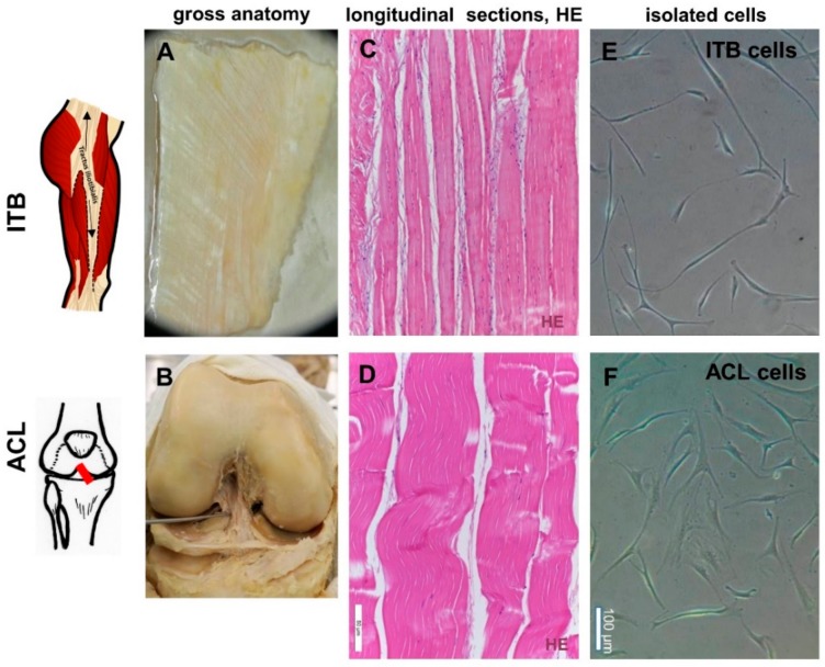
Figure 1.
The gross anatomy, histology, and isolated fibroblasts of iliotibial band (ITB) and anterior cruciate ligament (ACL) tissue: The gross anatomy of ITB (A) and ACL (B) tissue. The hematoxylin and eosin (HE) staining of ITB (C) and ACL (D) tissue. Fibroblasts isolated from ITB (E) and ACL (F) tissues. Scale bars: 50 µm (Figure 1C,D) and 100 µm (Figure 1E,F). ITB: iliotibial band, ACL: anterior cruciate ligament.

